

3D Image Segmentation of Brain Tumors Using Deep Learning
₹3,000.00 Exc Tax
Huge Price Drop : 50% Discount
Source Code + Demo Video
Description
ABSTRACT
Among brain tumors, gliomas are the most common and aggressive, leading to a very short life expectancy in their highest grade. Thus, treatment planning is a key stage to improve the quality of life of oncological patients. Magnetic Resonance Imaging (MRI) is a widely used imaging technique to assess these tumors, but the large amount of data produced by MRI prevents manual segmentation in a reasonable time, limiting the use of precise quantitative measurements in clinical practice. So, automatic and reliable segmentation methods are required; however, the large spatial and structural variability among brain tumors make automatic segmentation a challenging problem. In this paper, we propose an automatic segmentation method based on Convolutional Neural Networks (CNN), exploring small 3×3 kernels. The use of small kernels allows designing a deeper architecture, besides having a positive effect against overfitting, given the fewer number of weights in the network. We also investigated the use of intensity normalization as a pre-processing step, which though not common in CNN-based segmentation methods, proved together with data augmentation to be very effective for brain tumor segmentation in MRI images.
INTRODUCTION
Gliomas are the brain tumors with the highest mortality rate and prevalence. These neoplasms can be graded into Low-Grade Gliomas (LGG) and High Grade Gliomas (HGG), with the former being less aggressive and infiltrative than the latter. Additionally, the tumor mass effect changes the arrangement of the surrounding normal tissues. Also, MRI images may present some problems, such as intensity inhomogeneity, or different intensity ranges among the same sequences and acquisition scanners. In brain tumor segmentation, we find several methods that explicitly develop a parametric or non-parametric probabilistic model for the underlying data. Other methods are known as Deep Learning deal with representation learning by automatically learning a hierarchy of increasingly complex features directly from data. So, the focus is on designing architectures instead of developing handcrafted features, which may require specialized knowledge. CNN’s have been used to win several object recognition and biological image segmentation challenges. Since a CNN operates over patches using kernels, it has the advantages of taking context into account and being used with raw data.
PROPOSED METHOD
In this paper, we propose an automatic segmentation method based on Convolutional Neural Networks (CNN). Here, deep learning architecture is introduced in order to segment the brain tumor with a sufficient accuracy in a shorter time compared to the other basics methods. the results of the various steps on segmentation improvement showed 98% accuracy as compared to the basic algorithm.
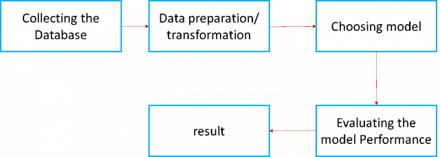
BLOCK DIAGRAM
CONCLUSION
In summary, we propose a novel CNN-based method for the segmentation of brain tumors in MRI images. Experimental results show that our model achieves better classification results in MRI image classification of tumors, and the accuracy of the test reached 98.55%.


Reviews
There are no reviews yet.