


Lung Cancer Detection using Matlab
₹3,000.00
Huge Price Drop : 50% Discount
Source Code + Demo Video
100 in stock
Description
ABSTRACT
Effective identification of carcinoma at AN initial stage is a vital and crucial facet of image process. many Segmentation strategies are accustomed observe carcinoma at early stage. during this paper, AN approach has been given which is able to diagnose carcinoma at AN initial stage exploitation CT scan pictures. one in all the key challenges is to get rid of white Gaussian noise from the CT scan image, that is completed exploitation Gabor filter and to phase the respiratory organ is rework technique twin tree complicated moving ridge rework (DTCWT) is employed. The GLCM options square measure extracted from the processed image to create feature vector. These options is compared with information pictures exploitation classifier as neural networks . when distinguishing the established un wellness as traditional or neoplasm we have a tendency to square measure segmenting the tumour image by exploitation watershed segmentation to induce colour options of tumour when obtaining colour options for form options we have a tendency to square measure applying FCM. during this paper BPNN square measure applied for the detection of carcinoma realize to search out the severity of un wellness and that we find completely different quality attributes like accuracy, sensitivity(recall), exactness and specificity to grasp the performance.
DEMO VIDEO
INTRODUCTION
Lung cancer is one in all the foremost common cancers, accounting for over 225,000 cases, 150,000 deaths, and $12 billion in health care prices yearly within the U.S its additionally one in all the deadliest cancers, overall, solely revolutionary organization 17 November of individuals within the U.s. diagnosed with carcinoma survive 5 years when the diagnosing, and also the survival rate is lower in developing countries . The date of a blight refers to about abundantly it’s metastasized. Stages one and a pair of talk to cancers localized to the lungs and latter stages talk to cancers that have unfold to alternative organs. Current diagnostic strategies embody biopsies and imaging, like CT scans. Early detection of carcinoma (detection throughout the sooner stages) considerably improves the possibilities for survival, however it’s additionally tougher to observe early stages of carcinoma as there square measure fewer symptoms. Our task may be a binary classification downside to observe the presence of carcinoma in patient CT scans of lungs with and while not early stage carcinoma. we have a tendency to aim to use use strategies from pc vision and deep learning, notably 2nd and 3D convolution neural networks, to make AN correct classifier. AN correct carcinoma classifier may speed up and cut back prices of carcinoma screening, granting a lot of widespread early detection and improved survival. The ambition is to assemble a computer-aided diagnosing (CAD) arrangement that takes as ascribe accommodating chest CT scans and outputs whether or not or the accommodating has carcinoma.
EXISTING SYSTEM
Watershed
The segmentation obtained from thresholding features a heap of noise- several voxels that were a part of respiratory organ tissue, particularly voxels at the sting of the respiratory organ, cared-for fall outside the vary of respiratory organ tissue radio density because of CT scan noise. this implies that our classifier won’t be ready to properly classify pictures during which cancerous nodules square measure settled at the sting of the respiratory organ.
Thresholding
Typical radio densities of varied components of a CT scan square measure shown in Table one. Air is usually around −1000 HU, respiratory organ tissue is usually around −500, water, blood, and alternative tissues square measure around zero HU, and bone is usually around 700 HU, thus we have a tendency to mask out pixels that square measure getting ready to −1000 or on top of −320 to go away respiratory organ tissue because the solely phase.
DRAWBACKS
- Database isn’t used
- If thresholding is low correct detection impossible
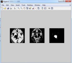
PROPOSED METHOD
In this, to get a lot of correct results, a tendency to divide the work into the subsequent 3 stages: Image Enhancement: to enhance the image and eliminate the noise, corruption or interference , 3 strategies square measure used: Gabor filter (has the most effective results), motorcar improvement algorithmic rule, and twin tree complicated moving ridge rework (DTCWT) is done.
Image Segmentation: To segregate and phase the improved pictures, the strategies used are: Thresholding approach and Marker-Controlled Watershed Segmentation approach (which offers higher results than thresholding).options Extraction stage: to get the precise options of the improved mesmeric image exploitation Binarization and Masking Approach.
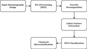
BLOCK DIAGRAM

ADVANTAGES
- Noise is reduced exploitation Gabor filter
- DTCWT is completed for eliminating high frequencies
- CNN as a classifier it checks a lot of inputs to classify
- By exploitation FCM we have a tendency to get form options of neoplasm
METHODOLOGIES
- CT pictures
- GLCM
- FCM
- DTCTWT
APPLICATIONS
- Bio medical
- Cancer detection and diagnoisis
SOFTWARE REQUIREMENTS
- Matlab 7.14 above
CONCLUSION
An image improvement technique is developing for earlier illness detection and treatment stages; the time issue was taken in account to get the abnormality problems in target pictures. Image superior and accurateness is that the amount factors of this analysis, angel superior appraisal still as aspartame date wherever were adopted on low preprocessing techniques accurate physicist clarify a part of Gaussian rules. The planned technique is economical for segmentation principles to be an area of interest foundation for feature extraction getting. The planned address offers awfully able after-effects assay with altered acclimated techniques. counting on accepted options, a course allegory is formed. the most detected options for correct pictures comparison square measure pixels proportion and mask-labeling with high accuracy and sturdy operation.
REFERENCES
[1] Anita chaudhary, SonitSukhraj Singh “Lung Cancer Detection on CT Images by Using Image Processing”2012 International Conference on Computing Sciences
[2] NihadMesanovic, HarisHuseinagic, Matija Males, , MislavGrgic, Emir Skejic, MuamerSmajlovic ”Automatic CT Image Segmentation of the Lungs with Region Growing Algorithm”
[3] SayaniNandy, Nikita Pandey “A Novel Approach of Cancerous Cells Detection from Lungs CT Scan Images’’ International Journal of Advanced Research in Computer Science and Software Engineering Volume 2, Issue 8, August 2012
[4] Prof. Samir Kumar Bandyopadhyay “Edge Detection From Ct Images Of Lung’’ International Journal Of Engineering Science & Advanced Technology Volume – 2, Issue – 1, 34 – 37
[5] FatmTaher, NaoufelWerghi and Hussain Al-Ahmad “Extraction of Sputum Cells using Thresholding Techniques for Lung Cancer Detection” 2012 International Conference on Innovations in Information Technology
[6] QinghuaJi,Ronggang Shi “A Noval Method of Image Segmentation Using Watershed Transformation”2011 International Conference on Computer Science and Network Technology
Additional information
| Weight | 0.000000 kg |
|---|

Reviews
There are no reviews yet.