


Liver Tumor Detection using Matlab
₹3,000.00
Huge Price Drop : 50% Discount
Source Code + Demo Video
100 in stock
Description
ABSTRACT
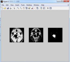
Now –a- days liver Cancer is growing anonymously in huge rate because this liver cancer has low survival rate even symptoms do not appear until the cancer in the advanced stage if disease caught late the average person survive only a year so we propose the cancer detection using clustering and Neural networks In this we go with the three phases of detection processing phase, preprocessing phase and detection phase where the wavelets are applied to signified the segmentation to classify the normal and abnormal stages of the tumor Accurate detection of size and location of brain tumor plays a vital role in the diagnosis of tumor. The diagnosis method consists of four stages, pre-processing of MR images, feature extraction, and classification. After histogram equalization of image, the features are extracted based on Discrete wavelet transformation (DWT). In the last stage, Probabilistic Neural Network(PNN) are employed to classify the Normal and abnormal brain. An efficient algorithm is proposed for tumor detection based on the K-Means Clustering.
INTRODUCTION
Liver cancer is the type of cancer that starts in liver it doesn’t spreads outside the area of the liver The liver, which is located below the right lung and under the ribcage, is one of the largest organs of the human body. It has a range of functions, including removing toxins from the body, and is crucial to survival. Since, we are going to deal with Liver cancer, we are concerned about the liver cancer diagnosis tests. There are various tests for diagnosis of liver cancer. Among these tests CT scan and MRI is of utmost important. Generally, every doctor ask for CT scan to look for the tumor in the liver. If they find the liver affected, then later on they ask for MRI to get detail knowledge about the tumor in the liver. Because MRI provides a better view and proper tumor location.
EXISTING SYSTEM
In existing system we used only CT scan images by that we cant find the microorganisms over the liver by thresholding we can find only the max and min values of segregation and for extracting the image used Discrete cosine transform and classified by Support vector machine the extraction not be in proper and accuracy low
PROPOSED METHOD
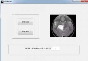
The proposed approach consists of four successive stages as shown in Figure Firstly, image enhancement stage, to improve the quality of the input image. The second stage, is image segmentation to extract the liver and tumor. The third stage is feature extraction and selection to extract the main features of the tumor object using special and transformation domains techniques. Finally, the probalistic neural network classifier is employed to classify the tumor as benign or malignant.
BLOCK DIAGRAM

ADVANTAGES
- High accuracy
- Low complexity
METHODOLOGIES
- DWT
- GLCM Feature Extraction
- PNN Training and Classification
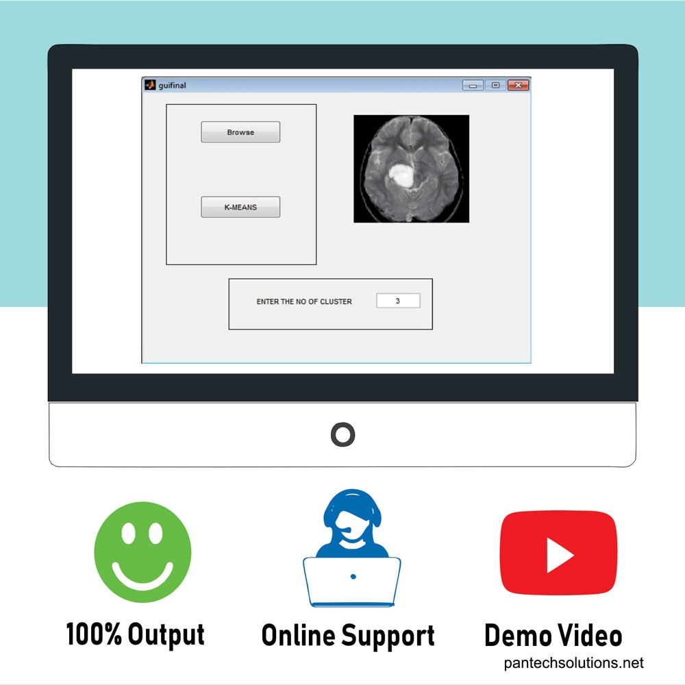
- K-Means Clustering
SOFTWARE REQUIREMENT
- MATLAB 7.14 above versions
REFERENCES
[1] Weimin Huang; Ning Li; Ziping Lin; Guang-Bin Huang; Weiwei Zong; Jiayin Zhou; Yuping Duan, “Liver tumor detection and segmentation using kernel-based extreme learning machine,” inEngineering in Medicine and Biology Society (EMBC), 2013 35th Annual International Conference of the IEEE , vol., no., pp.3662- 3665, 3-7 July 2013
[2] Jie Lu; Defeng Wang; Lin Shi; Pheng Ann Heng, “Automatic liver segmentation in CT images based on Support Vector Machine,” in Biomedical and Health Informatics (BHI), 2012 IEEE-EMBS International Conference on , vol., no., pp.333-336, 5-7 Jan. 2012
[3] R. K. Bhullar and N. K.Walia. A New Hybrid Technique for Detection of Liver Cancer on ltrasound Images. International Journal of Science and Research (IJSR), Vol. 3,No. 10, pp. 1647- 1651, India,2014
[4] V.v.gomathi and s.karthikeyan. Performance evaluation of hmsk and sqfd algorithms for computer tomography (ct) image segmentation of effective radiotherapy. Journal of Theoretical and Applied Information Technology, Vol. 22,No. 2, pp. 1647- 1651, India,2014
[5] R.Rajagopal and P.Subbiah. Computer Aided Detection of Liver Tumor using SVM Classifier. International Journal of Advanced Research in Electrical, Electronics and Instrumentation Engineering, Vol. 3,No. 6, pp. 10170- 10177, India, June 2014.
[6] B. V. Ramana1, Prof. M.Surendra Prasad Babu, and Prof. N. B. Venkateswarlu. A Critical Study of Selected Classification Algorithms for Liver Disease Diagnosis (IJAREEIE). International Journal of Database Management Systems ( IJDMS ), Vol. 3,No. 2, pp. 101- 114, India, May 2011
[7] Li Ma; Yang, L., “Liver Segmentation Based on Expectation Maximization and Morphological Filters in CT Images,” in Bioinformatics and Biomedical Engineering, 2007. ICBBE 2007. The 1st International Conference on , vol., no., pp.690-693, 6-8 July 2007
[8] Yoshida, H.; Keserci, B.; Casalino, D.D.; Coskun, A.; Ozturk, O.; Savranlar, A., “Segmentation of liver tumors in ultrasound images based on scale-space analysis of the continuous wavelet transform,” in Ultrasonics Symposium, 1998. Proceedings., 1998 IEEE , vol.2, no., pp.1713-1716 vol.2, 1998
DEMO VIDEO
Reviews
There are no reviews yet.