

Leukocytes Classification and Segmentation in Microscopic Blood Smear
₹3,000.00
Huge Price Drop : 50% Discount
Source Code + Demo Video
100 in stock
Description
ABSTRACT
Analyzing digital magnifier pictures for earlier Acute Myelogenous Leukemia (AML) designation and treatment need refined computer code and hardware systems. during this paper presents hand-picked mathematical ways used for image segmentation and application of moving ridge remodel for the subsequent segments classification by multi-resolution decomposition of segments corpuscle pictures. The Haar moving ridges remodel and Daubechies wavelet remodel approach has been adopted here and used for feature extraction permitting its use for image denoising and backbone sweetening furthermore. Feature classification is then achieved by self-organizing neural networks. A planned methodology has been verified for simulated structures and so offers the higher segmentation accuracy and exactitude for analysis of microscopic pictures.
INTRODUCTION
Microscopic pictures of the blood cells area unit discovered to seek out out several diseases. Changes within the blood condition show the event of diseases in a private. leukemia will result in death if it’s left untreated. supported some statistics it’s found that the cancer of the blood is that the fifth reason behind death in men and sixth reason behind death in girls. leukemia originates within the bone marrow. every bone contains a skinny material within it that is additionally called a bone marrow. The parts of blood area unit Red Blood Cells (erythrocytes), White Blood Cells (leukocytes), platelets and plasma. leukemia is detected solely by analyzing the white blood cells. therefore our study is targeted solely on the white blood cells (WBCs). The cells within the bone marrow begin dynamic and that they get infected and become leukemia or infected cells.
EXISTING SYSTEM
K-means agglomeration it clusters the image and signifies the blood cells and classifies with Support vector machine, however, it’s a time taking method by coaching the set
PROPOSED SYSTEM
In a manual methodology of leukemia detection, specialists check the microscopic pictures. this can be prolonged and time taking method that depends on the person’s ability and not having a customary accuracy. leukemia detection system analyses the microscopic image and overcomes these drawbacks. It extracts the desired elements of the photographs and applies some filtering techniques. Segmentation and agglomeration approach is employed for white blood cells detection and it classifies and trains with NN it shows the suitable quantitative relation of the parameters with none information loss
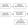
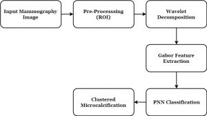
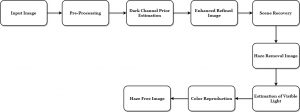
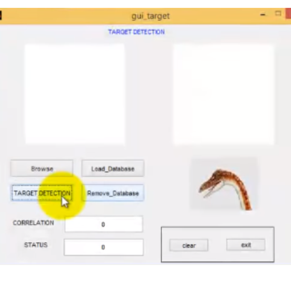
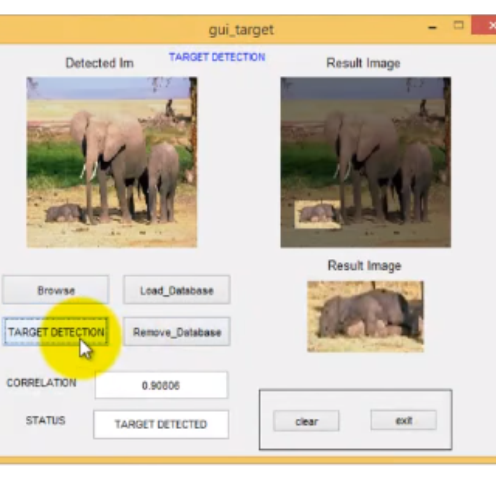
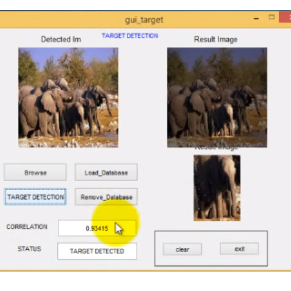
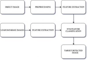
BLOCK DIAGRAM

DEMO VIDEO
APPLICATIONS
- Medical Imaging
- Bio-Medical
CONCLUSION
The major a part of this work is to section the lymphocytes and myelocytes white blood cells for leukemia detection. the primary part of the planned system is coping with the image cleansing and noise removal for creating the image prepared for the any and correct study. The second and major part is that the leucocytes identification from the image. The third part is coping with the nucleus and protoplasm extraction from the image which may finally be used for the feature extraction within the last part of the planned system. This model has been tested against thirty-three pictures taken below the same lightning condition and therefore the accuracy achieved is ninety-three .57% [19]. we will additionally use the planned system to seek out out the proportion of leukemia infection in the microscopic image. we tend to hope this approach is going to be useful for today’s quick life and early detection of leukemia with none would like of pricey tests and with a much better accuracy
FUTURE WORK
The future work may be implemented with alternative algorithms associated with this method once understand used methodologies have some constraints. any analysis can specialize in an assortment of a lot of samples to yield higher performance Associate in Nursing building an overall system for cancer classification.
Related products
-
-
Sale!
Add to cart

-

-

-
Sale!


Reviews
There are no reviews yet.