


Brain Pressure Analysis using Neural Networks
₹3,000.00
Huge Price Drop : 50% Discount
Source Code + Demo Video
99 in stock
Description
ABSTRACT
Automatic defects detection in MR images is very important in many diagnostic and therapeutic applications. Because of high quantity data in MR images and blurred boundaries, tumour segmentation and classification is very hard. This work has introduced one automatic brain tumour detection method to increase the accuracy and yield and decrease the diagnosis time. The goal is classifying the tissues to three classes of normal, begin and malignant.. In MR images, the amount of data is too much for manual interpretation and analysis. During past few years, brain tumor segmentation in magnetic resonance imaging (MRI) has become an emergent research area in the field of medical imaging system. Accurate detection of size and location of brain tumor plays a vital role in the diagnosis of tumor. The diagnosis method consists of four stages, pre-processing of MR images, feature extraction, and classification. After histogram equalization of image, the features are extracted based on Dual-Tree Complex wavelet transformation (DTCWT). In the last stage, Back Propagation Neural Network(BPN) are employed to classify the Normal and abnormal brain. An efficient algorithm is proposed for tumor detection based onthe Spatial Fuzzy C-Means Clustering.
INTRODUCTION
Tumors are two types one is primary tumor and second one is secondary tumor. The tumor cell is present within skull and grows within skull is called primary tumor. Malignant tumors are primary tumors. The tumor presents outside the skull and enter into the skull region called secondary tumor. Metastatic tumors are examples of secondary tumors [1]. The tumor takes up place in the skull and interferes with the normal functioning of the brain. Tumor shifts the brain towards skull and increases the pressure on the brain. Detection of tumor is the first step in the treatment. A tumor is an intracranial solid neoplasm or abnormal growth of cells within the brain and lung or the central spinal canal. Tumor is one of the most Common and deadly diseases in the world. Detection of the tumor in its early stage is the key of its cure. There are many different types of tumors that make the decision very complicated. So classification of tumor is very important, in order to classify which type of tumor really suffered by patient. A good classification process leads to the right decision and provide good and right treatment. Treatments of various types of tumor are mostly depending on types of tumor. Treatment may different for each type, and usually Brain contains more number of cells that are interconnected to one another different cells control different parts of the body. Some cells control the leg movement. likewise others cells of the brain controls other parts in the body. Tumors may have different types of symptoms ranging from headache to stroke, so symptoms will vary depending on tumor location .Different location of tumor causes different functioning disorder.
The general symptoms of tumor
- Headache in early mornings.
- Gradually loss of movement in leg.
- Loss of sensation in arm.
- Loss of vision in one or both eyes.
- Speech difficulty.
Magnetic Resonance Imaging (MRI) is widely used in the scanning. The quality of image is high in the MRI. The quality of image is main important in tumor. MRI provides an unparalleled view inside the human body In MRI we can see detailed information exordinarly compared to any other scanning like X-ray, C.T scans. The contrast of tumor cell is high compared to normal brain cell. Treatments techniques for the tumor
- Surgery
- Radiation therapy
- Chemotherapy
EXISTING SYSTEM
- Thresholding method
- C means clustering
- Manual analysis – time consuming, inaccurate and requires intensive trained person to avoid diagnostic errors.
DRAWBACKS
- Difficult to get accurate results
- Not applicable for multiple images for Tumor detection in a short time
- Medical Resonance images contain a noise caused by operator performance which can lead to serious inaccuracies classification.
PROPOSED SYSTEM
- Neural Network for classification
- K-means clustering for tumor detection and structural analysis
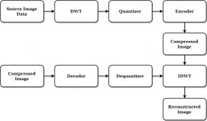
BLOCK DIAGRAM

METHODOLOGIES
- DWT
- GLCM Feature Extraction
- NN Training and Classification
- K-Means
ADVANTAGES
- It can segment the Brain regions from the image accurately.
- It is useful to classify the Brain Tumor images for accurate detection.
- Brain Tumor will be detected in an early stages
APPLICATION
- Brain Tumor diagnosis system for medical application
SOFTWARE REQUIREMENTS
- MATLAB 7.14 and above versions
CONCLUSION
In this paper the tumor detection and classification is successfully implemented by a novel algorithm for Tumor Classification is presented. This new method is a combination of discrete wavelet Transform and convolution Neural Network along with the implementation of GLCM. By using these algorithms an efficient Tumor Classification method was constructed with maximum recognition rate Simulation results using Tumor database demonstrated the ability of the proposed method for optimal feature extraction and efficient Tumor classification. The ability of our proposed Tumor Classification method is demonstrated on the basis of obtained results on Tumor image database. On other Tumor image databases the other combinations are there for training and test samples.
REFERENCES
1] S. Bauer et al., “A survey of mri-based medical image analysis for brain tumor studies,” Physics in medicine and biology, vol. 58, no. 13, pp. 97– 129, 2013.
[2] D. N. Louis et al., “The 2007 who classification of tumours of the central nervous system,” Acta neuropathologica, vol. 114, no. 2, pp. 97–109, 2007.
[3] E. G. Van Meir et al., “Exciting new advances in neuro-oncology: The avenue to a cure for malignant glioma,” CA: a cancer journal for clinicians, vol. 60, no. 3, pp. 166–193, 2010.
[4] G. Tabatabai et al., “Molecular diagnostics of gliomas: the clinical perspective,” Acta neuropathologica, vol. 120, no. 5, pp. 585–592, 2010.
[5] B. Menze et al., “The multimodal brain tumor image segmentation benchmark (brats),” IEEE Transactions on Medical Imaging, vol. 34, no. 10, pp. 1993–2024, 2015.
[6] N. J. Tustison et al., “N4itk: improved n3 bias correction,” IEEE Transactions on Medical Imaging, vol. 29, no. 6, pp. 1310–1320, 2010.
[7] L. G. Ny´ul, J. K. Udupa, and X. Zhang, “New variants of a method of mri scale standardization,” IEEE Transactions
DEMO VIDEO

Reviews
There are no reviews yet.